

Pathologies and Procedures
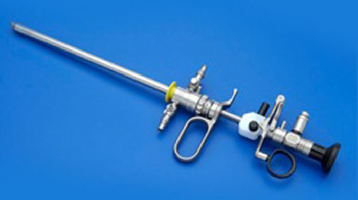
Hysteroscopy
Diagnostic hysteroscopy is a minimally invasive endoscopic technique used to visualize the uterine cavity. A thin instrument called a hysteroscope is introduced through the vagina (usually without the need for a speculum) and passes through the cervical canal. Once inside the uterus, it enables a complete examination of the uterine cavity. Saline solution is infused through the hysteroscope to expand the cavity and allow for detailed assessment.
This procedure typically does not require anesthesia and is usually painless.
Diagnostic hysteroscopy can identify conditions such as:
- Endometritis (inflammation of the endometrium)
- Endometrial hyperplasia (thickening of the endometrium)
- Uterine malformations (septate or bicornuate uterus)
- Synechiae (intrauterine adhesions)
- Polyps
- Myomas
- Neoplasms
Polypectomy is often the first operative hysteroscopic procedure performed during hysteroscopy training. It
It involves the removal of cervical or endometrial polyps, with the latter requiring hysteroscopic resection under direct vision for complete removal – unlike curettage, which has a high failure rate.
No pharmacological preparation is needed. In reproductive-age women, the ideal time for the procedure is during the proliferative phase of the menstrual cycle.
The resection technique typically involves slicing the polyp from the top down to the base until the root is severed. In some cases, if the polyp is pedunculated and not too large, it can be removed in one piece.
A myoma is a benign fibromuscular mass that can be solitary or multiple. It may develop on the outer surface, within the uterine wall, or protrude into the cavity if deeply embedded. Its size can vary significantly.
Myomectomy – the surgical removal of one or more fibroids (multiple myomectomy) – is the most conservative surgical approach. It aims to restore the anatomical and functional integrity of the uterus.
Fibroids, also called myomas, develop a capsule around them, clearly separating them from the surrounding tissue, which can facilitate removal. However, the degree of intramural involvement in submucosal fibroid is the main limiting factor of this technique. Greater intramural involvement increases surgical difficulty, complication risk and the chance of incomplete removal.
To guide treatment, fibroids are classified into three types:
G0 myomas (Type 0) protrude completely into the endometrial cavity without any intramural extension. For this reason, they are the easiest to treat regardless of their size, with the highest success rate and the lowest risk of complications.
Submucosal myomas that predominantly extend into the endometrial cavity but have an intramural extension of less than 50% of their total volume are classified as Type 1 (G1).
Submucosal myomas that protrude less than half of their volume into the endometrial cavity and have an intramural extension greater than 50% are classified as Type 2 (G2).
In any case, the thickness of the myometrium between the outer margin of the myoma and the serosal surface of the uterus should not be less than 5 mm. This thickness, known as the myometrial free margin, is measured via ultrasound during the patient’s preoperative evaluation. An increased degree of intramural extension leads to a longer surgical procedure, greater technical difficulties, a higher risk of incomplete removal of the fibroid tissue, the risk of intravasation (the passage of the uterine distension fluid into the patient’s intravascular compartment), as well as the risk of uterine perforation and hemorrhage.
In some cases, preoperative administration of GnRH analogs may be necessary, as they reduce the volume and vascularization of the myoma, making its removal easier and safer.
Uterine malformations are usually congenital anomalies. They often do not cause significant problems, but in approximately 3% of cases, they can lead to infertility or recurrent miscarriages.
One such malformation is the presence of a fibrous median septum, which may partially or completely divide the uterine cavity and, rarely, extend into the vagina.
Hysteroscopic surgery can correct this malformation and restore normal uterine anatomy and function through a procedure called metroplasty.
Synechiae are adhesions between the walls of an organ cavity, resulting from previous trauma or inflammatory events. They can be endouterine or endocervical, forming for example after procedures involving the uterine cavity such as curettage, scraping or VTP (voluntary termination of pregnancy), hysteroscopic surgery.
Synechiae can vary in severity and present different symptoms. They often cause hypomenorrhea (scant menstrual flow) or, if particularly extensive and firm, amenorrhea (total absence of menstruation). On their own, they can lead to infertility by causing stenosis of the cervical canal or uterine cavity, thereby preventing sperm progression or implantation of the fertilized egg.
Laparoscopy
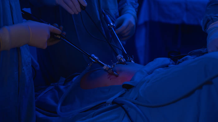
If the patient desires pregnancy but is experiencing difficulty conceiving, laparoscopy may help the physician identify – and sometimes correct – the underlying pathology.
Through endoscopic visualization, the surgeon can verify whether the Fallopian tubes (through which the fertilized egg passes) are open or not. The condition and patency of the fimbriae, which are the distal portions of the tube responsible for capturing the egg, can also only be accurately assessed through laparoscopy. During laparoscopic evaluation, in addition to the direct visualization of all pelvic organs involved in reproductive function (uterus, tubes, ovaries, peritoneum), certain procedures are routinely performed, while others are executed only if necessary.
In particular, chromosalpingoscopy is always performed. It involves the injection of a colored dye (methylene blue) through the cervical canal and the vaginal route.
The filling of the tubes and the dye’s passage through the fimbriae into the peritoneum are observed. It is practically a test for tubal patency, but not for functionality. In extreme cases, in women already candidates for reproductive medicine techniques (IVF/ICSI), where the pathology has irreversibly compromised tubal function, there may be a clear indication for removing one or both tubes (salpingectomy).
Conservative Treatment
- Ovarian biopsy
- Vaporization treatment with Diode or CO2 Laser
- Vaporization treatment with Argon
- Vaporization treatment with Plasma-jet
- Enucleation of ovarian or tubal cysts
- Enucleation of para-ovarian or para-tubal cysts
- Conservative removal of ectopic pregnancy
- Removal of ovarian fibroids
- Lysis of peri-ovarian or peri-tubal adhesions
- Treatment of hydrosalpinx or PID
- Drainage or removal of tubo-ovarian abscess
Destructive Treatment
- Oophorectomy
- Salpingectomy
- Salpingectomy for ectopic pregnancy
- Annexectomy
Endometriosis is one of the most important indications for surgical laparoscopy. Cases of minimal, mild, moderate, or severe endometriosis – even those involving endometriotic cysts or large nodules – can be brilliantly treated. Endometriosis is a disease that affects millions of women of reproductive age (3-10%) worldwide. It is rare before menarche and tends to decrease significantly after menopause. The term derives from “endometrium,” the tissue that lines the inner surface of the uterus and is shed each month during menstruation.
In endometriosis, tissue similar to the endometrium is found outside the uterus in other areas of the body. In these locations, endometrial tissue develops as superficial lesions, cysts, or deep infiltrating nodules, most commonly in the pelvis, affecting the ovaries, tubes, uterine ligaments, the anatomical area located laterally and/or dorsally to the uterus (parametrium), the area between the vagina and the rectum, the intestinal wall (sigmoid-rectum), the urinary tract (ureters), the bladder wall, or the peritoneum.
The most common extra-pelvic locations include the inguinal canal, small intestine, appendix, ileocecal valve, right colon, diaphragm, pleura, lungs, abdominal wall (from previous surgeries).
The most frequent symptoms in cases of endometriosis include:
- Pain before and during menstruation (dysmenorrhea)
- Pain during sexual intercourse (dyspareunia)
- Pain during defecation (dyschezia)
- Diarrhea and/or constipation
- Painful urination (dysuria)
- Chronic pelvic pain
- Bleeding in stools (hematochezia/rectorrhagia)
- Blood in urine (hematuria)
- Nausea/vomiting
- Irregular menstrual cycles
Some women with endometriosis may also have no symptoms. Infertility affects approximately 30-40% of women with endometriosis and is a common outcome as the disease progresses. The severity of the pain is not necessarily related to the extent or size of the lesions. Small formations (called petechial) often produce more prostaglandins -substances that activate inflammatory processes- than larger lesions. This may explain why small implants are sometimes more symptomatic.
Symptoms often tend to intensify over time, although in some cases, there are periods of remission and recurrence.
Endometriosis always requires staging, which can be pre-operative (by expert and dedicated sonographers or radiologists) or intraoperatively.
There are various classification systems.
The most well-known is the rAFS (Revised American Fertility Society) staging system, which divides the disease into 4 stages.
However, this does not correlate the stage with the clinical complexity of the disease (especially for deep infiltrating forms) and is no longer used in reference centers.
The most complete and widely used staging systems today are the #Enzian Score and AAGL 2021.
S
A myoma is a benign mass, either singular or multiple, of fibromuscular tissue that can develop on the outer surface or within the thickness of the uterus, reaching the uterine cavity when it is very deep. Its size can also be considerable.
Myomectomy, the removal of one or more fibroids (multiple myomectomy), is the most conservative surgical procedure for treating this condition and aims to restore the anatomical and functional integrity of the uterus. Fibroids (or fibromas, similar terms) grow by creating a capsule on their surface that clearly delineates them from the surrounding tissue and often allows for easy removal. The fibroid nodules are then removed from the abdominal cavity using a special instrument called a “morcellator,” which shreds the fibroids, making their removal possible through small skin access points. This procedure may sometimes require a larger skin incision (up to 2 cm). In cases of myomectomy for large nodules, a higher incision (a few centimeters above the navel) is necessary.
The myoma is always extracted from the abdominal cavity using a morcellation system performed inside a bag (endo-bag) to avoid the risks of dissemination and contamination.
It is the size, number, and sometimes the location of the fibroids that determine the difficulty of the procedure. This type of surgery is characterized by varying times, obviously depending on the complexity of the individual cases.
With laparoscopic hysterectomy, the entire uterus (and sometimes the ovaries and tubes) can be completely removed, even if it is large. The uterine manipulator used for laparoscopic hysterectomy is different from the traditional one. It is larger and has a series of silicone valves to complete the disconnection of the uterus without losing gas pressure within the abdomen, which would otherwise leak through the vagina. Once the uterus is laparoscopically disconnected, it is removed vaginally or through a bag (endo-bag), and the vaginal dome is closed with a continuous suture. Laparoscopic hysterectomy can also be associated with procedures to repair pelvic floor defects (cystocele, urethrocele, rectocele) or surgeries to prevent vaginal dome prolapse. In cases of hysterectomy for large uterus, a higher incision (a few centimeters above the navel) is necessary.
Peritoneal adhesions are one of the main causes (12%) of sub-occlusive and occlusive abdominal pain syndromes, which often require multiple surgeries and have a significant socio-economic impact due to the increased incidence of morbidity and mortality. The increase in patients with adhesion syndrome is also related to the increase in life expectancy and survival after resective surgeries for abdominal neoplasms. In the past, symptomatic adhesion syndrome was treated through laparotomy. A new surgical incision on the patient’s abdomen would once again trigger the pathophysiological mechanism responsible for adhesion formation. The spread of laparoscopic techniques and the refinement of its therapeutic possibilities, along with the positive results achieved, have led us to reconsider the clinical issue of adhesion syndrome in its various aspects. Looking through the laparoscopic lens into the peritoneal cavity of a person with chronic abdominal pain, utilizing this technique, which serves both as diagnostic and therapeutic, means improving the patient’s quality of life. Laparoscopic results are excellent and, in most cases, allow complete liberation of the pelvis.
Endoscopica Malzoni is the only center in Italy affiliated with Adhesion Related Disorder (ARD) Education and Awareness, a society that selects and coordinates worldwide the most qualified centers for the treatment of pathologies related to adhesions (www.adhesionrelateddisorder.com). Sometimes, it may be necessary to make an incision two fingers below the left costal arch along its midline (Palmer’s point). Then, using a mini-optic (2 mm), the presence of adhesions below the navel is checked, and only under visual control, the optical trocar is inserted.
Involuntary urine loss (incontinence), objectively demonstrable, is a personal and social issue. By having the patient cough several times with a full bladder, one can determine whether incontinence truly exists. In these cases, it is referred to as stress incontinence or incontinence due to exertion. This should be distinguished from another type of incontinence due to hyperactivity of the detrusor muscle of the bladder, called urge incontinence. Urge incontinence is the most common form in older women. Urge incontinence benefits from pharmacological and behavioral treatments. Stress incontinence, especially when associated with pelvic organ prolapse, requires surgical treatment.
Until a few years ago, the classic treatment for stress incontinence was a procedure known as plastic surgery (anterior colporrhaphy); this surgery, still widely practiced, has a low long-term success rate. Mixed forms of urinary incontinence (stress incontinence combined with urge incontinence) should be studied on a case-by-case basis to determine the most effective and safe therapy.
Other surgical options include:
- Vaginal Sling surgery (TVT, TOT, TVT-Secure, etc.)
- Laparoscopic promontofixation for uterine or vaginal dome prolapse
- Laparoscopic colposuspension according to McCall
- Laparoscopic repair of lateral cystocele defect (paravaginal repair)
- Laparoscopic repair of rectocele
Laparoscopy is increasingly used in the oncological field at reference centers. The possibility of performing lymphadenectomy using the sentinel lymph node technique (the first lymph node station potentially affected by neoplastic pathology) or systematic pelvic and/or para-aortic lymphadenectomy through a mini-invasive technique allows for the treatment of early-stage cervical cancer and endometrial carcinoma, possibly associated with extrafascial or radical hysterectomy. Surgical staging of ovarian cancer (early stage) or evaluation of cytoreductibility in advanced stages is also possible. The surgical results obtained in terms of the radicality of the intervention, from an oncological standpoint, are considered, at reference centers, fully comparable to those of traditional surgery.
In addition to the common benefits of laparoscopy, patients also gain an oncological advantage, as they are generally able to begin potential adjuvant treatments (chemotherapy or radiotherapy) very early.
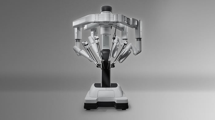
At centers affiliated with Malzoni Research Hospital, it is possible to perform robotic surgery procedures with the assistance of the Da Vinci Xi Robot, in collaboration with the physicians at the Advanced Pelvic Surgery Center.
Our division routinely performs vaginal surgeries or those using the V-NOTES system. The main indications include:
- Uterine prolapse (hysterocele)
- Cystocele or cystourethrocele (bladder prolapse)
- Stress urinary incontinence due to urethral hypermobility
- Urge urinary incontinence
- Rectocele (rectal prolapse)
- Cervical hypertrophy
- Bartholin’s gland cyst
Possible interventions include:
- Vaginal hysterectomy with or without removal of ovaries/tubes
- Repair of fascial support structures in the anterior and/or posterior compartments with or without mesh (prosthetic mesh)
- Placement of Sling (prosthetic bands) (TVT, TVT-O, TVS)
- Tracheloplasty or trachelectomy
- Marsupialization or excision of Bartholin’s cyst