

Laparoscopy
Introduction
Laparoscopy is a modern, well-established surgical technique that is now considered the Gold Standard in the treatment of gynecological diseases.
The Malzoni Research Hospital has been using this method since 1995 for the treatment of all gynecological conditions. Laparoscopy is a specialist surgical procedure that allows gynecological treatments to be performed without opening the abdominal wall, using a few small incisions on the skin.
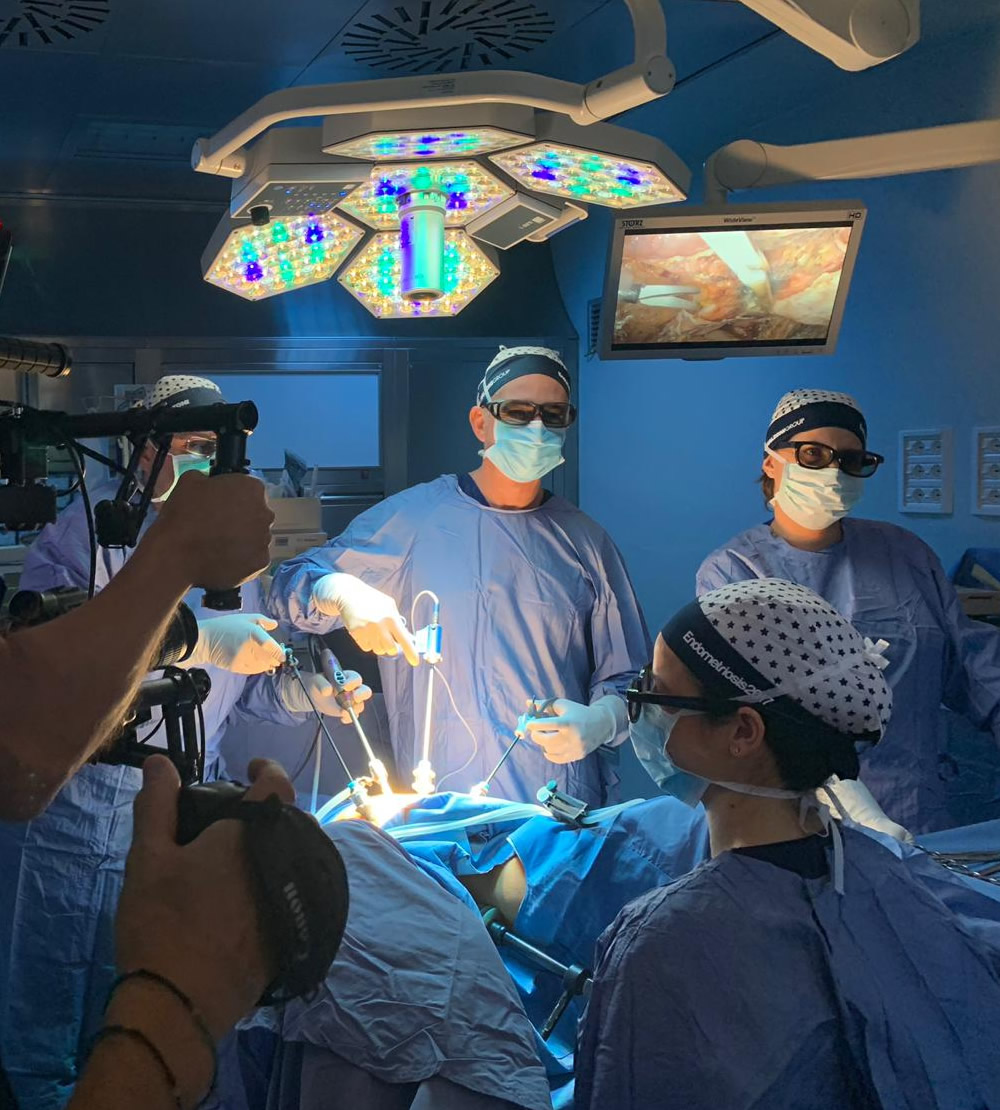
Thanks to technological advancements and the miniaturization of instruments, the surgical team can perform the procedure while observing images on a monitor, using 2 to 3 very thin instruments inserted through small incisions in the lower abdomen, allowing the surgical steps to be carried out.
At the end of the procedure, the skin access points will be closed with sutures (usually absorbable) that do not require removal.
Historical Background
Laparoscopic surgery in gynecology was born in Europe in 1940, thanks to Palmer, who used endoscopy to examine the fallopian tubes in cases of infertility.
Initially, this technique was used exclusively for diagnostic purposes to avoid unnecessary exploratory laparotomies, for example, to diagnose suspected appendicopathy or ectopic pregnancy.
In the 1970s, the German school of Semm (1970) and the French school of Bruhat and Manhes opened new therapeutic possibilities for endoscopy.
The first procedures were performed for adnexal pathology (ovarian cysts, adhesions, and tubal occlusions) and ectopic pregnancies. As surgical instrumentation improved and suturing techniques developed, the range of possible procedures expanded.
In 1979, Kurt Semm described the first laparoscopic myomectomy: after the mechanical enucleation of the fibroid, sutures were applied. In the United States, in 1989, Harry Reich performed the first fully laparoscopic hysterectomy.
Surgical Technique
The procedure is performed under general anesthesia (with orotracheal intubation) using thin surgical instruments and an optic (camera) that are inserted into the abdomen through micro-incisions ranging in diameter from 5 to 12 mm. The procedure always requires the distension of the abdominal cavity with a gaseous mixture of CO2, which is rapidly reabsorbed at the end of the operation. The duration of the procedure varies depending on the complexity of the anatomical region involved. As is routine for all surgical procedures, a venous access is placed for the administration of necessary medications.
Once sedation has been achieved, a urinary catheter is also placed, which is generally removed immediately postoperatively before waking up, unless there is a need to keep it for specific clinical requirements. At the end of the procedure, it may be necessary to place an abdominal drain and/or a nasogastric tube, which will be removed in the following days. For certain procedures involving the urinary tract, the placement of a ureteral stent (unilateral or bilateral) may be required—an internal device that keeps the ureter open and is therefore not visible externally, to be removed within variable timeframes depending on clinical needs.
Very rarely, blood/plasma transfusions may be necessary. Although the risk is extremely low, our facility is equipped with a Postoperative Intensive Care Unit (T.I.P.O.) with dedicated anesthesiology and nursing staff available 24/7. The resumption of normal postoperative feeding depends on the physiological recovery of intestinal activity.
This is generally very early unless the surgical procedure directly involves intestinal loops, in which case a slightly longer interval may be required. When clinically permissible, early mobilization is encouraged to facilitate a faster postoperative recovery. Typically, the average length of hospital stay from admission to discharge is 48-72 hours, though it may extend to 7 days or more for more complex surgeries (intestine, urinary tract, bladder).
A moderate tenderness at the site of skin scars is common, usually short-lived and manageable with common painkillers. A frequent occurrence is also pain in the clavicular area, related to phrenic nerve irritation, which is also temporary. Postoperatively, the patient may experience paresthesia/hypoesthesia, i.e., subjective sensations of mild altered skin sensitivity (a feeling of “numbness,” tingling, warmth), particularly in the abdominal area or lower limbs (inner thigh). These effects are generally due to the position assumed during surgery and tend to resolve spontaneously within a few days in most cases, though more rarely they may persist for a few weeks.
Our facility also has a pathology service for intraoperative histological examination, although the final evaluation is still subject to verification of the definitive histological results. Our operating rooms are equipped with state-of-the-art instrumentation fully dedicated to advanced endoscopic surgery and also feature 3D surgical vision devices, providing additional advantages in terms of visualization and, consequently, greater accuracy of the surgical procedure.
Advantages and Complications
Laparoscopy offers significant advantages over traditional surgery.
LAPAROSCOPY (a minimally invasive surgical technique) is currently recognized as the “gold standard” (treatment of choice) for most gynecological procedures due to its undeniable advantages over laparotomic techniques (traditional open-abdomen surgery):
- Magnified vision with greater surgical precision
- Reduced blood loss
- Lower risk of postoperative adhesions
- Less postoperative pain
- Faster recovery and shorter hospital stay
- Better aesthetic results
Like all medical and surgical procedures, laparoscopy can also involve risks.
In our case studies, the risk of intra- and postoperative complications is low. The risks associated with the procedure may be general, specifically related to laparoscopic technique, or to the surgical procedure itself (depending on the affected area/organ).
- Infectious
- Hemorrhagic
- Thrombotic
- Anesthesiologic
General risks, as with all surgical procedures, are also negatively influenced by any concomitant conditions the patient may have. Among these, the most common are infectious and hemorrhagic complications.
Fever in the immediate postoperative period is common and may be associated with the inflammatory process typically linked to the surgical procedure. If persistent, it may indicate an underlying infectious process requiring further diagnostic investigations (instrumental and laboratory tests) to determine its origin. Generally, it resolves with the administration of appropriate antibiotic and anti-inflammatory therapy, with reoperation being rarely necessary. The risk of intraoperative hemorrhage is extremely low, and if it occurs, it is promptly managed during surgery. Even when it happens, it is usually controlled while maintaining a laparoscopic approach, with conversion to laparotomy being required in only very rare cases (<0.01%). The risk of postoperative abdominal bleeding is slightly higher but still extremely low. Often, these are self-limiting events, though in some cases, depending on severity, they may require blood transfusions along with other medical treatments. The need for reoperation is rare. A slightly more frequent but still minimal risk is the formation of an organized hematoma or bruise within the abdominal wall near the surgical scars. These cases are generally self-limiting but, depending on their extent, may sometimes require percutaneous drainage along with medical therapy. The need for reoperation remains rare. As with all surgical procedures, patients should also be informed of the extremely remote risk of fatality associated with the procedure, which remains exceedingly rare even in the case of postoperative complications.
- Injury to a major arterial or venous vessel
- Intestinal injury and subsequent peritonitis
- Gas embolism
- Abdominal wall emphysema
- Pneumothorax
- Incisional hernias
- Cellular dissemination from non-benign pathologies
The extraction of tissues and/or organs suspected of malignancy (e.g., highly cellular fibroids, large-volume uterus, suspicious adnexal cysts) is performed using specialized devices (dedicated bags or endobags of appropriate size). This procedure prevents the dissemination of potential neoplastic cells since surgical maneuvers (including fragmentation or morcellation of fibroids or the uterus) are carried out inside the bag, which protects and isolates the peritoneal surfaces.
- Intra- or postoperative hemorrhage requiring transfusion (very low <1%)
- Ureteral injury (<1%)
- Bladder injury (1%)
- Intestinal perforation (<2%)
- Intestinal-vaginal, vesico-vaginal, or intestino-vesical fistulas (2%)
- Vaginal suture dehiscence (4%)
- Neurological damage (<1%)
- Infection, erosion or migration of Mesh implants
For ureteral injury, this refers to a disruption in the continuity of the ureter wall, possibly leading to urine leakage into the abdomen, often associated with peritonitis symptoms. It may result from thermal or mechanical damage. Radiological contrast imaging may sometimes be required for confirmation. In some cases, it can be resolved simply by placing a ureteral stent, which must remain in place for several weeks or months; otherwise, a laparoscopic or laparotomic reoperation is needed for surgical cleansing and suturing or resection (with reanastomosis or reimplantation) of the affected segment. Even in this case, a ureteral stent is necessary, and depending on the situation, a bladder catheter may also be required for a few weeks. For bladder injury, this refers to a disruption in the continuity of the bladder wall, which may result in urine leakage into the abdomen and is often associated with peritonitis symptoms. It may be caused by thermal or mechanical damage. In some cases, radiological contrast imaging may be required for confirmation. Occasionally, small lesions may heal spontaneously with prolonged bladder catheterization for several weeks; otherwise, a laparoscopic or laparotomic reoperation is necessary for surgical cleansing and suturing of the affected segment. In this case, a bladder catheter must also remain in place for a few weeks.
Intestinal perforation may occur in most cases within a few days after surgery. It consists of a wall lesion with possible leakage of intestinal contents into the abdomen, often associated with peritonitis symptoms. It is generally related to excessive distension of intestinal loops and may result from thermal or mechanical damage. A laparoscopic or laparotomic reoperation is required for surgical cleansing and suturing of the affected segment, if possible, often with the need for a protective stoma.
A fistula is the formation of an abnormal communication pathway between two adjacent but normally separate organs. It can occur between the intestine and the vagina, the bladder and the vagina, or, much more rarely, between the intestine and the bladder. This phenomenon, although rare, may develop in the immediate postoperative period or later. It may result from thermal, mechanical, infectious, or inflammatory damage. In the case of an intestinal fistula, air and/or fecal material may be observed passing from the bladder or the vagina. In the case of a vesico-vaginal fistula, urinary leakage may be observed from the vagina. Radiological contrast imaging may sometimes be required for confirmation. The resolution is generally surgical, but several weeks may be needed for complete healing.
Vaginal suture dehiscence refers to the breakdown of the vaginal closure performed after a hysterectomy. This phenomenon may occur spontaneously, sometimes due to infectious complications, or mechanically, often following sexual intercourse—especially if resumed earlier than the recommended three-month period. If the lesion is small, it may heal spontaneously with medical therapy; otherwise, surgical repair via vaginal access or, more rarely, laparoscopic access is necessary.
Neurological damage. Permanent neurological injuries are rare but more likely in cases of parametrial surgery due to the dense innervation of this anatomical area. Despite always using nerve-sparing techniques to minimize the possibility of nerve structure injury, nerve preservation is not always possible, especially if affected by pathology (e.g., endometriosis).
For the risk of neurological damage, the potential role of post-surgical fibrosis must also be considered in patients who have undergone multiple surgeries. This fibrosis, by altering normal anatomical planes, makes any subsequent surgery even more complex and risky.
The Mesh (synthetic grafts) used for the correction of uro-genital prolapse may rarely become infected, eroded, or migrate. If the postoperative complication does not resolve completely with medical therapy, an additional surgical procedure may sometimes be required for partial or complete removal of the mesh.